
What Is the Mitral Valve Area Calculator?
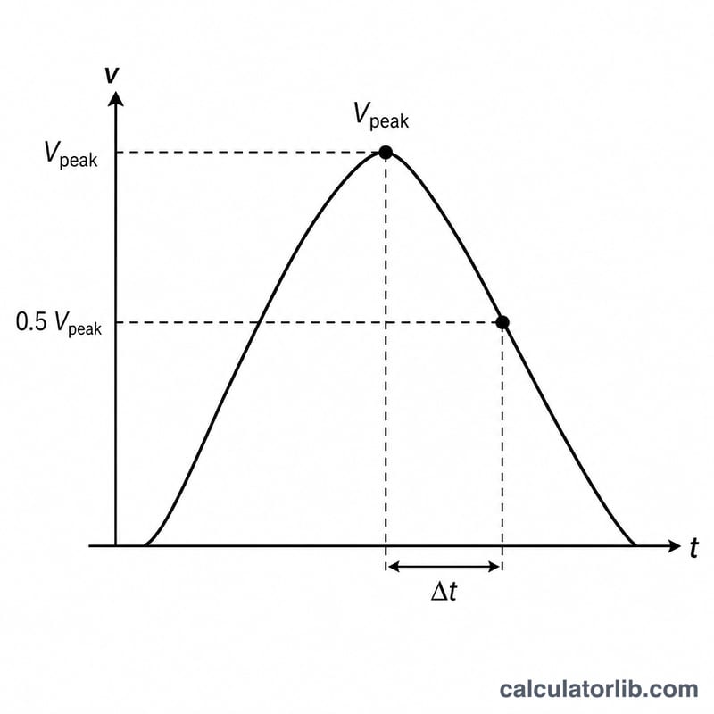
This calculator estimates the mitral valve area (MVA) from the Doppler-derived pressure half-time (PHT) measured during echocardiography. The pressure half-time is the time it takes for the peak transmitral pressure gradient to fall to half its initial value. In patients with mitral stenosis, a tighter (smaller) valve area slows pressure decay, lengthening the PHT. The relationship was popularized by Hatle and colleagues and remains a routine clinical estimate.

How to Use It
Enter the pressure half-time in milliseconds (ms) as measured from the mitral inflow Doppler tracing. The calculator divides the empirical constant 220 by your PHT value to return the estimated valve area in square centimeters (cm²). A larger PHT yields a smaller estimated valve area.
The Formula Explained
The equation is simply $$\text{MVA} = \frac{220}{\text{Pressure Half-Time (ms)}}$$ where 220 is an empirically derived constant and PHT is in milliseconds. For example, a PHT of 220 ms gives an area of exactly 1.0 cm². Mitral stenosis is generally graded as mild (MVA > 1.5 cm²), moderate (1.0–1.5 cm²) and severe (< 1.0 cm²).
Worked Example
Suppose a patient has a measured pressure half-time of 440 ms. Then $$\text{MVA} = \frac{220}{440} = 0.5 \text{ cm}^2$$ indicating severe mitral stenosis. A PHT of 110 ms would give \(\frac{220}{110} = 2.0\) cm², a normal-to-mild range.
FAQ
Is this a diagnostic tool? No. It is an educational estimate. Clinical decisions must rely on a full echocardiographic assessment by a qualified cardiologist.
When is the PHT method unreliable? It can be inaccurate immediately after balloon valvuloplasty, with significant aortic regurgitation, abnormal LV/LA compliance, or tachycardia. In these settings other methods (planimetry, continuity equation) are preferred.
Why the constant 220? It is an empirical value derived from clinical correlation studies relating PHT to invasively measured valve areas.