What this calculator does
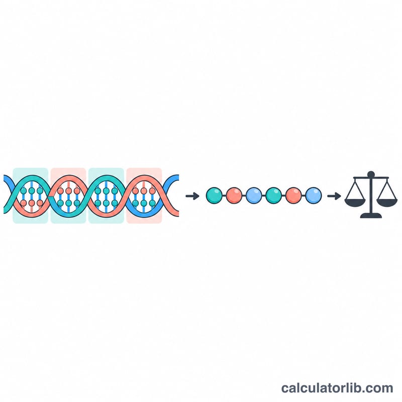
This tool reads a DNA coding sequence (5′→3′), translates it with the standard genetic code in the reading frame you choose, and calculates the average molecular weight of the resulting protein. It accepts plain sequences or FASTA text, treats U as T, and ignores whitespace and digits. Knowing the mass in kDa helps you plan SDS-PAGE gels, choose purification columns, estimate extinction and yield, and check that an expressed construct matches the expected band size.
How to use it
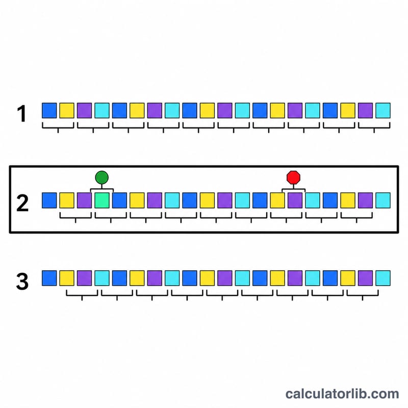
Paste your coding sequence, pick frame 1, 2 or 3, and decide whether translation should stop at the first stop codon (typical for a single gene) or skip stops and continue. The result shows the weight in kDa and Da, the number of amino acids, GC content and the translated protein sequence.
The formula explained
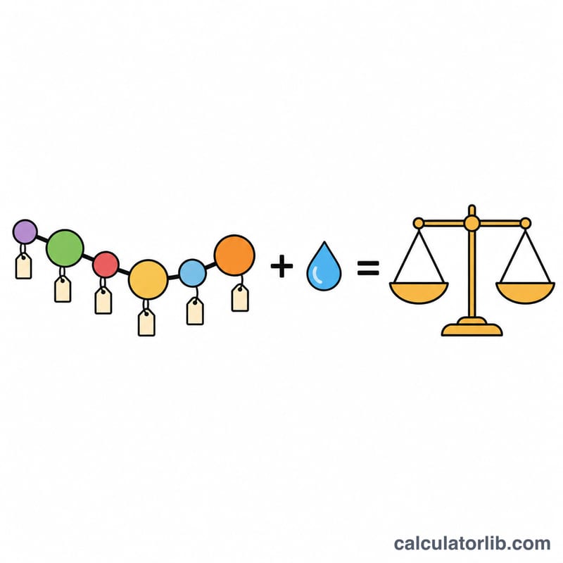
Each amino acid contributes its residue mass, which is the free amino acid mass minus one water lost in the peptide bond. One water is added back for the N-terminal H and C-terminal OH.
$$\text{MW} = \sum_i n_i \, m_i + 18.01524$$Here \(n_i\) is the count of amino acid \(i\) and \(m_i\) is its average residue mass in Da. Then \(\text{kDa} = \text{Da}/1000\).
Worked example
Take ATGGGCTAA in frame 1. It reads ATG (Met), GGC (Gly), TAA (stop), so the protein is Met-Gly.
$$\text{MW} = 131.1926 + 57.0519 + 18.0152 = 206.2597\,\text{Da} = 0.2063\,\text{kDa}$$Amino Acid Average Residue Masses
The calculator sums the average (isotope-weighted) mass of each amino acid residue, i.e. the amino acid minus one water lost on peptide-bond formation, then adds one water (18.01524 Da) for the free N-terminus (H) and C-terminus (OH) of the whole chain. Values are in daltons (Da).
| Amino acid | 3-letter | 1-letter | Average residue mass (Da) |
|---|---|---|---|
| Glycine | Gly | G | 57.0519 |
| Alanine | Ala | A | 71.0788 |
| Serine | Ser | S | 87.0782 |
| Proline | Pro | P | 97.1167 |
| Valine | Val | V | 99.1326 |
| Threonine | Thr | T | 101.1051 |
| Cysteine | Cys | C | 103.1388 |
| Leucine | Leu | L | 113.1594 |
| Isoleucine | Ile | I | 113.1594 |
| Asparagine | Asn | N | 114.1038 |
| Aspartic acid | Asp | D | 115.0886 |
| Glutamine | Gln | Q | 128.1307 |
| Lysine | Lys | K | 128.1741 |
| Glutamic acid | Glu | E | 129.1155 |
| Methionine | Met | M | 131.1926 |
| Histidine | His | H | 137.1411 |
| Phenylalanine | Phe | F | 147.1766 |
| Arginine | Arg | R | 156.1875 |
| Tyrosine | Tyr | Y | 163.1760 |
| Tryptophan | Trp | W | 186.2132 |
Water added once per chain: 18.01524 Da.
Example by hand: the dipeptide Met-Gly has mass \(131.1926 + 57.0519 + 18.01524 = 206.25974\) Da.
Standard Genetic Code Codon Table
This is the standard (universal) nuclear genetic code, grouped by amino acid. Codons are written as DNA (A, C, G, T); if your sequence is RNA, U is read as T. ATG (Met) is the usual start codon, and TAA, TAG and TGA are the stop codons. Some organelles (e.g. mitochondria) and a few organisms use variant codes, which this standard table does not cover.
| Amino acid | Codons | Number |
|---|---|---|
| Phe (F) | TTT, TTC | 2 |
| Leu (L) | TTA, TTG, CTT, CTC, CTA, CTG | 6 |
| Ile (I) | ATT, ATC, ATA | 3 |
| Met (M) | ATG (start codon) | 1 |
| Val (V) | GTT, GTC, GTA, GTG | 4 |
| Ser (S) | TCT, TCC, TCA, TCG, AGT, AGC | 6 |
| Pro (P) | CCT, CCC, CCA, CCG | 4 |
| Thr (T) | ACT, ACC, ACA, ACG | 4 |
| Ala (A) | GCT, GCC, GCA, GCG | 4 |
| Tyr (Y) | TAT, TAC | 2 |
| His (H) | CAT, CAC | 2 |
| Gln (Q) | CAA, CAG | 2 |
| Asn (N) | AAT, AAC | 2 |
| Lys (K) | AAA, AAG | 2 |
| Asp (D) | GAT, GAC | 2 |
| Glu (E) | GAA, GAG | 2 |
| Cys (C) | TGT, TGC | 2 |
| Trp (W) | TGG | 1 |
| Arg (R) | CGT, CGC, CGA, CGG, AGA, AGG | 6 |
| Gly (G) | GGT, GGC, GGA, GGG | 4 |
| Stop | TAA, TAG, TGA | 3 |
Total: 61 sense codons + 3 stop codons = 64.
Interpreting Your Protein Mass
The result is the theoretical average mass of the unmodified polypeptide encoded by your sequence. 1 kDa = 1000 Da, so a result of 33,000 Da is 33 kDa. As a rough sanity check, the average amino acid contributes about 110 Da, so \(\text{MW (Da)} \approx N_{\text{aa}} \times 110\): a 100-residue protein is about 11 kDa and a 300-residue protein about 33 kDa. Real proteins deviate from this estimate depending on their composition.
Why the SDS-PAGE band may differ from the calculated mass
- Tags and fusions: a His6 tag adds roughly 0.8 kDa of residues plus any linker; larger fusion partners add much more. Check whether your sequence includes the tag and whether you translated it.
- Signal peptide cleavage: secreted or membrane proteins often lose an N-terminal signal sequence (commonly around 15–30 residues), so the mature protein is lighter than the full translation.
- Post-translational modifications: glycosylation, phosphorylation, lipidation, proteolytic processing, removal of the initiator Met and disulfide bonds (about −2.016 Da each) change the true mass. Glycosylation can add several kDa and smear a band.
- Wrong frame or strand: a different reading frame gives a completely different protein, often ending early at a stop codon. If the result is far too small, try frames 1–3 and consider the reverse complement of your sequence.
- Gel behavior: SDS-PAGE is only approximate (often within about 10%); highly acidic, basic or hydrophobic proteins can migrate anomalously.
- Stop codons: how an internal or terminal stop is handled (set by the stop codon option) changes how many residues are counted.
Average vs monoisotopic mass
This calculator uses average masses, which weight each element by its natural isotope abundance. They correspond to the centroid of the isotope envelope and suit larger proteins in mass spectrometry. Monoisotopic mass uses only the most abundant isotope of each element (e.g. 12C) and is typically used for peptides and high-resolution instruments; it is slightly lower than the average mass, and the difference grows with size. Do not compare the two directly.
This tool is intended for research and planning of expression and purification experiments; it provides general information only, not medical or diagnostic advice.
FAQ
Does it include tags or modifications? No. Only the translated amino acids are counted, so add the mass of any tag not in your sequence, and note that phosphorylation, glycosylation or signal peptide cleavage change the observed size.
Why is my mass wrong? Check the reading frame and that your sequence is the coding strand, not the reverse complement.
Is the mass monoisotopic? No, it is the average mass, which matches gel and SEC estimates better than monoisotopic values.